Reports
Health
Wellness
Depression or major depressive disorder is a mental health condition that affects an individual's behavior, mood, and overall health.
People with depression may experience prolonged feelings of sadness, hopelessness, and emptiness.
Depression usually begins in early adolescence or adulthood but can appear at any age.
Depression is one of the most common mental health conditions in the United States.
It is also more common in women and people assigned female at birth (AFAB) than men and people assigned male at birth (AMAB).
Common symptoms of depression are:
The symptoms of depression may vary slightly in the younger age group.
Common depression symptoms in adolescents and teens are:
Though the exact cause of depression is unclear, the disorder runs in families, hinting that genetic factors may increase one's risk of developing the condition.
Research is still underway to study the genetic variations that increase the risk for depression.
Other factors that may increase one’s risk for depression include:
Genetics plays a central role in the development of depression.
Scientists believe that nearly 40% of people with depression have a genetic link, while environmental and other factors make up for the remaining 60%.
Existing research states that a complex interplay of genes and other environmental (non-genetic) factors determine an individual’s risk of developing depression.
Few studies have reported that people having a first-degree relative with depression (a parent, sibling, or child) are three times more likely to develop the condition than the general population.
This shows that depression runs in families, and the disease has a significant hereditary component.
Some people can also develop the condition without a family history.
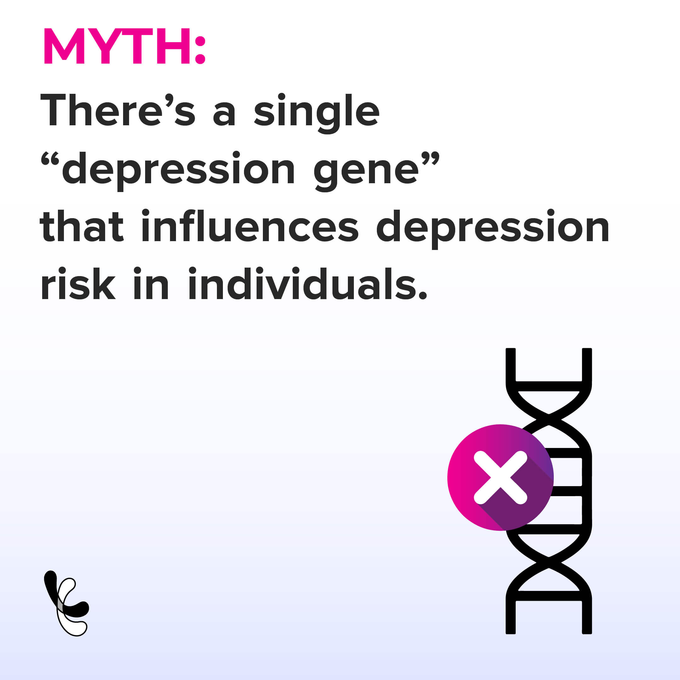
Though genetics influences the development of depression and anxiety, no single causative gene has been identified.
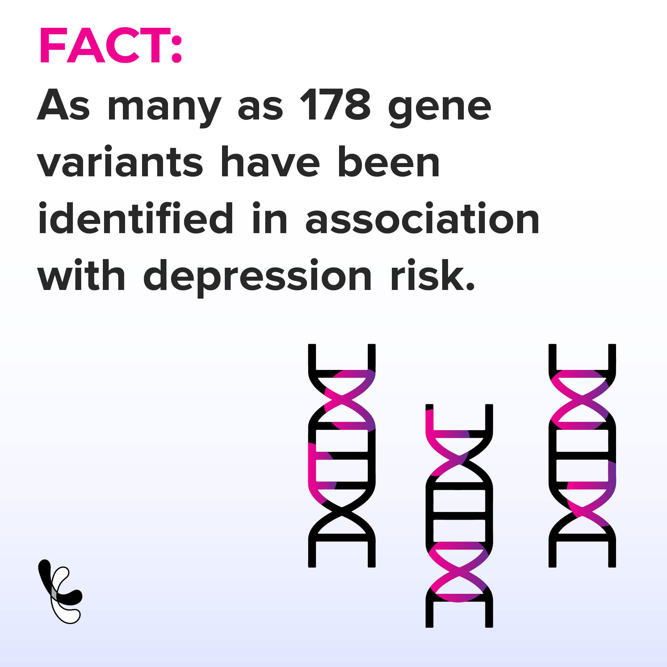
Research states that genetic variations on multiple genes contribute to the development of depression.
However, having a genetic variant does not confirm you will develop the condition.
Besides genetic variations, how genes are passed down to the next generation (mode of inheritance) may also affect an individual’s risk for depression.


A study published in The American Journal of Psychiatry reported that the research team isolated a gene prevalent in several families with a history of depression.
Chromosome 3p25-26 was found in over 800 families with recurrent depression.
Standard treatments used for depression are:
Most people require a combination of two or more treatments.
Did you know that your genes influence how well a treatment works for you? Several studies show that genes affect how your body absorbs, uses, and eliminates drugs like antidepressants.
Many genes influencing drug metabolism have been studied and are of specific interest to doctors and researchers.
If you have depression, it is essential to understand that treatment for this condition may take time.
You may have to undergo a couple of treatments to determine the most suitable one.
If you experience one or more symptoms of depression, it is important to seek medical care.
Before starting any antidepressant medications, informing your doctor about your medical history and current medication and supplements is essential.
This information will help your doctor determine the best treatment for you.
Learning about the genetic background of depression can help understand the condition better and provide optimal treatment options.
Engaging in regular physical activities can help weight management and strengthen our bones and muscles. In addition, it can help us stay healthy and free from diseases like cardiometabolic disorders, diabetes, and hypertension. A recent study has reported when older adults stay active, their brain releases more of a class of proteins that lead to better cognition.
Our brain helps us respond to external signals by accepting and transferring the signs through our brain cells.
The brain cells communicate at a specialized junction called the synapse.
They are tiny gaps between the preceding brain cell that sends the information and the succeeding cell that receives it.
Moving your body and indulging in regular physical activities is good for our physique and our brain.
In addition, physical activity can boost our cognition - the ability to think, respond, and solve problems.
Exercises can also help us reduce the risk of cognitive decline.
Research suggests that inactive adults have twice the risk of cognitive decline than active adults.
According to the Physical Activity Guidelines for Americans, adults need to perform at least two types of physical activity every week to maintain their physical and mental health.
The researchers at the Weill Institute for Neurosciences and the University of British Columbia reported that exercise improves brain aging in older adults.
The study is the first to use human data to show that physical activity is linked to synaptic proteins and better cognition.
Synapses are connection points between brain cells that help maintain cognition. They are composed of proteins that facilitate the transmission of information.
The researchers obtained the data from the Memory and Aging Project at Rush University in Chicago and tracked the physical activity of the elderly participants during their senescence.
The participants have also agreed to contribute their brains after they die.
The findings were coherent with previous findings that people who had more of these proteins when they died had better cognition in their late life.
Exercise and body movements support and stimulate the normal functioning of proteins, resulting in transmitting signals throughout the brain.
It is believed that amyloids and tau (proteins involved in Alzheimer's) disintegrate the brain cells.
However, research suggests that synaptic integrity (a measure of healthy neuronal transmission) measured in the brain tissues of autopsied adults weakens the relationship between Alzheimer's proteins, leading to brain degeneration.
Elderly adults with higher levels of proteins associated with synaptic integrity have a reduced risk of Alzheimer's disease.
Thus, sustaining healthy synaptic integrity can protect our brain from neurodegenerative diseases, which can be maintained by performing physical activities and exercises.
Get A Comprehensive Fitness Analysis with the Gene Fitness Report
https://www.sciencedaily.com/releases/2022/01/220107100955.htm
https://www.cdc.gov/nccdphp/dnpao/features/physical-activity-brain-health/index.html
https://www.ncbi.nlm.nih.gov/pmc/articles/PMC6770965/
Get Deep Fitness Insights From Your 23andMe, AncestryDNA Raw Data
Excess sugar in the body or hyperglycemia is a prevalent condition affecting almost 3.5 million people in the US (according to a 2017 report). The Van Andel Research Institute study reports how an excess intake of dietary carbohydrates can damage mitochondrial integrity and, thereby, function.
According to the CDC, in 2017-18, the dietary sugar intake of Americans was 17 teaspoons. However, the American Heart Association (AHA) only recommends six teaspoons per day. Sugar is broken down (metabolized) in the body to glucose.
Now, the regulation of glucose levels in the body is closely controlled. Any imbalance in the glucose level leads to metabolic disorders, most commonly, diabetes.
Hyperglycemia occurs when the blood glucose concentration rises above 140 mg/dl (7.8 mmol/l).
Excess glucose levels can lead to insulin resistance, where the cells in your muscles, fat, and liver do not respond to insulin. When this happens, the blood glucose levels begin to rise. Persistent insulin resistance ultimately leads to type 2 diabetes.
A mitochondrion is an organelle in the body and is popularly known as the powerhouse of the cell. Mitochondria (plural) are the centers where energy production occurs. They supply energy to all cells and the whole body.
Adipose tissues are the primary controller of whole-body energy regulation. They store excess energy (in the form of triglycerides) and dissolve the same to provide power to the body in the event of energy scarcity.
There are two types of adipose tissues: brown adipose tissue and white adipose tissue.
WAT is responsible for storing excess energy, and BAT expends energy at times of energy scarcity. Beige adipocytes (adipose or fat cells) are at a basic level WAT cells. But, at times of extreme duress, beige adipose cells act as BAT and provide the body with extra energy.
Reportedly, mitochondrial malfunction in adipose cells contributes to metabolic disorders, predominantly obesity, and type 2 diabetes. Thus, one of the main causative factors of metabolic disorders is defective cell metabolism - which purportedly is caused by flawed energy production and distribution.
Additionally, a mitochondrial malfunction has adverse effects on the body.
Scientists at the Van Andel Research Institute explored the effects of excess sugar intake on mitochondrial integrity and function.
Scientists used a genetically altered mice model to allow the cells to accept excess sugar intake. Mitochondrial function was then measured using BAT thermogenesis (the process of heat production by brown adipose tissue).
The following were observed in the study:
PUFA or polyunsaturated fatty acids are important for supporting mitochondrial functioning and other biological processes like inflammation, cell to cell communication, and blood pressure regulation.
Saturated fatty acids are what are popularly known as bad fats. This fatty acid isn't nearly as useful or flexible as the PUFA. This disturbs the lipid composition of the mitochondrial membrane, thereby damaging the mitochondrial integrity.
Restoring normal glucose levels resulted in the balance of the fatty acid levels. This restored the lipid composition as well as the mitochondrial integrity and function.
The study found that the cellular changes brought on by a sugar-rich diet may go undetected or may not manifest under normal conditions; upon introduction of cellular stress (changes in cells that are inflicted by environmental stresses like extreme temperatures, exposure to toxins, etc.), these changes may become evident as functional deficiencies.
In conclusion, excess sugar intake throws off the lipid balance in the body. If the balance is not restored for a while, it can damage our mitochondria, making us more prone to an array of metabolic disorders like type 2 diabetes.
According to a research study by the University of Exeter Medical School in the United Kingdom, men with hemochromatosis, a common genetic disorder due to iron build-up, are ten times more likely to develop liver cancer.
Hemochromatosis, also called the iron-overload disease, is a condition where too much iron builds up in the body. Usually, the intestines absorb adequate amounts of iron and excrete the rest.
With hemochromatosis, excess iron is absorbed by the intestines, and the body has no way of getting rid of it. As a result, iron gets built up in joints, the pituitary gland, and organs like the liver, heart, and pancreas.
This gradually results in the shutting down of these organs if hemochromatosis is not treated.
Hemochromatosis is more serious in men. Women may be partially protected as they lose some iron during menstruation and childbirth.
Some common symptoms associated with hemochromatosis include:
HFE gene is associated with iron homeostasis. A variant (type) of the HFE gene, called the C282Y (the faulty type), is significantly associated with hereditary hemochromatosis.
According to a study published in the American Journal of Human Genetics, the C282Y variant contributes to 26% variation in ferritin levels among monozygotic twins.
With hemochromatosis, the iron build-up is commonly seen in the liver. This enlarges the liver and messes up the liver enzymes. It can result in an increased risk of liver conditions like cirrhosis, fibrosis, and cancer.
Hepatocellular carcinoma (HCC), a primary form of liver cancer, was the first condition in which hepatic iron overload was shown to predispose to the development of HCC.
According to a study, 8-10% of people with hemochromatosis develop HCC.
The study was led by the University of Exeter Medical School along with the University of Connecticut, Western University in Ontario, and South Warwickshire NHS Foundation Trust.
This study focused on men and women with two copies of the faulty HFE gene - C282Y. The data of 2890 people aged 40-70 years were analyzed over a nine-year period.
The following were observed:
The study insists on the importance of early diagnosis of hemochromatosis in order to avoid health complications and even death.
The NHS advises that “it is important to talk to your GP if you have a parent or sibling with hemochromatosis, even if you don’t have symptoms yourself” to identify your risk.
The lack of impact on women from the faulty HFE gene variant may suggest that periodic blood donations might play a protective role.
