Reports
Health
Wellness
It is normal for hair to lose color and turn gray as people age. However, today’s stressful lifestyle can accelerate this process resulting in grey hair appearing on people even in their 20s! Initially thought as a permanent change, new research reveals hair greying can be reversed - at least temporarily!
Hair greying occurs when melanin, a natural skin pigment that is responsible for hair, skin, and eye color, is gradually lost due to aging. At first, hair grows unpigmented out of the hair follicles.
Two pigments, melanin (produced by melanocytes) and keratin (produced by keratinocytes), together add color to the hair. Both melanocytes and keratinocytes are present near the follicles.
Melanin has two different versions - eumelanin and pheomelanin. Eumelanin gives rise to a red/yellow pigment (seen in blonde/ginger hair), and pheomelanin gives rise to a brown/black pigment (seen in brunette/black hair).
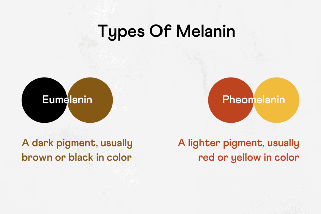
The distribution of eumelanin and pheomelanin is influenced by genetics, especially the MC1R gene.
As our body ages, so do stem cells - the parent cells of all functional cells of the body. Melanocytes are produced by melanocyte stem cells (McSCs).
Aging causes a loss of McSCs - so melanocyte production also gradually decreases. In the absence of melanin pigment, the hair turns grey.
Hair greying is influenced by genetic factors and the environment (including lifestyle). One of the major environmental contributors happens to be stress.
A previous study done by Harvard University explored stress-induced hair greying in mice. They observed that stress triggered the release of a chemical, norepinephrine, which affected the McSCs.
Norepinephrine accelerated the conversion of McScs to melanocytes, thereby depleting the population of McScs. This ultimately resulted in a complete halt in the hair coloration process.
This process was also noted to be irreversible in mice.
In 2016, researchers at UCL, UK, found the first-ever gene, IRF4 (interferon regulatory factor 4), associated with hair greying. IRF4 is actively involved in the regulation of the production and storage of melanin.
This gene interacts with MITF (microphthalmia-associated transcription factor) to activate tyrosine, a critical factor in melanogenesis (the process of melanin production). MITF has been known to repress McSCs survival and affect hair greying.
So, researchers hypothesize that IRF4 might influence the hair greying process. However, the exact mechanism behind this is still unclear.
Researchers at the Columbia University Irving Medical center undertook a small-scale study to observe how stress affected hair greying. The study included 14 participants from varying age groups, ethnicities, and sexes - most people, however, were white. The researchers collected dark, white, and two-colored hair (hair with partial greying) from these people.
The researchers used a digital and mathematical model to map and measure small color changes in a single hair. They found that over a stress-filled period, the hair started greying.
Surprisingly, the removal of the stress stimulus resulted in an apparent reversal of greying, and the hair became colored again.
To understand the mechanism behind this, the researchers studied thousands of other proteins in the hair. They observed changes in 300 out of the 1000 proteins as the hair greyed.
They also suspect the role of mitochondria in stress-induced hair greying. Mitochondria, other than being involved in energy production, also plays an important role in transmitting signals that respond to psychological stress.
A 70-year old with a head full of white hair cannot reverse hair greying by reducing stress. At the same time, 10-year-olds, no matter how much stress they experience, are not going to suddenly wake up to a head full of white hair.
The study reports a "threshold" or a limiting situation at which a hair turns grey permanently. Somewhere in middle age, as we approach this threshold, stress can accelerate the transition.
Reducing stress in life is most definitely a good thing. But once you cross this threshold, it's not going to reverse your hair greying.
When we are born, we inherit (receive) the genes from our parents, but we don't inherit their age.
In other words, how do older parents give rise to young offspring?
Scientists have tried to explain this by a process called "natural rejuvenation event." This event sets our biological age to zero, which is the beginning of aging in mammals.
This event happens as the zygote develops to form an embryo. A zygote is a single-celled organism resulting from a fertilized egg. The zygote goes through a cellular multiplication process to give rise to the multi-celled embryo.

Researchers are studying the possible application of this "age reversal event" as a therapeutic intervention for age-related diseases, such as arthritis or Parkinson’s.
Aging is something we all experience but actually know very little about. Hair greying, wrinkles formation, joint changes, etc., are signs of aging. Aging, on the other hand, encompasses all the processes that occur in the body that result in these signs - it is a combination of physiological changes in our bodies and environmental factors.
Aging eventually leads to physical denaturation, dysfunction, and ultimately mortality (death).
As we age, every cell in our body gets old due to the progressive accumulation of damage through the intervening years. But this cellular damage does not get passed on to human offspring to the next generation – the absence of this cellular damage in human offspring has baffled scientists for generations.
That's because germline cells (sex cells - egg and sperm that form the embryo/zygote) rejuvenate in the offspring after conception.
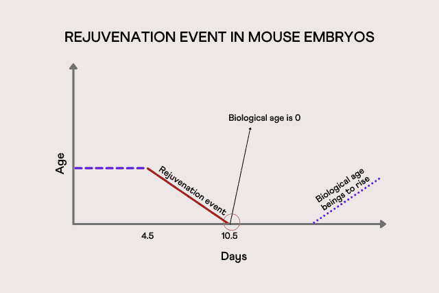
During the initial stages of embryogenesis (the formation of the embryo), life hits a reset button — the rejuvenation event in the embryo. When an embryo is attached to the uterus, the rejuvenation event sets the developing embryo to grow at its youngest biological age – zero. This rejuvenation of the embryo marks the beginning of the mammalian age.
The epigenetic aging clock is a biochemical test used to measure age. It was developed with conserved cytosines - units in DNA whose methylation levels change with age. Experts use these epigenetic clocks to predict the approximate ages of mouse embryos at their early stages of development.
They found that the age of mouse embryos stayed constant for the first few days after fertilization. However, by around 6.5 to 7.5 days into development, the biological age of the embryos witnessed a dip, indicating that they are undergoing some type of rejuvenation event.
The researchers say that the biological age of mice is set to zero, somewhere between 4.5 to 10.5 days after fertilization.
Eventually, during development, the biological age begins to slowly rise. However, the point exactly at which this growth happens is still unclear.
At present, similar data for humans is unavailable as studying human embryos at such early stages is prohibited. Recent studies have shown that the epigenetic clocks in cord blood can be used to estimate gestational age at birth.
When embryos can reset the biological clock, there is a hope to reverse the errors that occur due to cell damage, restoring the cells back to their agile state, eliminating signs of aging.
This hypothesis could possibly help researchers develop treatments for age-related diseases, such as arthritis or Parkinson’s - A paradigm shift in treating age-related illnesses.
Vitamin D deficiency is a common health problem worldwide among all age groups. Previous research studies have reported that ultraviolet (UV) exposure causes the skin to produce the hormone endorphin, which is chemically related to morphine, heroin, and other opioids.
A subsequent study reported that UV exposure raises endorphin levels in mice, displaying behavior consistent with opioid addiction. Recent research suggests that vitamin D supplementation may play a role in fighting opioid addiction.
Vitamin D, essential for a range of bodily functions, is predominantly obtained from exposure to sunlight. Once the body takes up vitamin D, it needs to be converted into its active form. Vitamin D is also naturally present in dietary sources like fish liver oils, fortified grains, dairy products, and egg yolks.
Vitamin D deficiency can occur when a person’s skin has an impaired ability to synthesize vitamin D from the sun. Individuals can also become deficient when their bodies cannot absorb the vitamin or convert it to its active form. People who avoid sun exposure, adhere to a vegan diet, or suffer from milk allergies may also be at a higher risk of vitamin D deficiency.
Vitamin D is vital for strong bones as it helps the body use calcium from the diet. Vitamin D deficiency has been associated with rickets, a condition leading to soft bones and skeletal deformities. However, increasing research suggests the importance of vitamin D in protecting against other health problems.
Know more: Genetic Factors That Could Increase Your Risk For Vitamin D Deficiency
Opioids are a form of narcotic prescribed as an over-the-counter pain medication. If incorrectly used, they can have serious side effects and even result in opioid addiction.
Individuals may gain tolerance to opioid medication if taken regularly and might need an increased dose to achieve the same effect in easing pain. They may also develop a dependence on the medication due to extended period use. In this case, individuals who abruptly stop the drug use may experience withdrawal symptoms.
A study led by Dr. David Fisher and his team of researchers at the Massachusetts General Hospital examined the potential link between UV radiation, vitamin D, and opioids.
Initially, the researchers began by studying two data sets.
The researchers drew the following conclusions from the data sets:
These results accounted for factors like age, gender, history of bone fractures, and chronic pain. However, further investigation was required to understand the association between low vitamin D and opioid use.
The researchers used mouse models to understand the patterns seen in the clinical data.
They induced vitamin D deficiency in the mice using the following methods:
50% of the diet-deficient mice were put on a regular diet for eight weeks before examining their response to morphine.
All the mice were then subject to a conditioned place preference (CCP) test in which the mice are placed in a multi-compartment chamber, trained to anticipate morphine in one chamber. Researchers then measured how long the mice spent inside the morphine chamber.
The following were observed:
The team found that the effect was reversed when they restored vitamin D levels using supplements. The researchers confirmed the results with a new batch of mice and additional sensory cues.
Furthermore, the researchers wanted to know whether vitamin D deficiency influenced how mice respond to opioids. For this, they placed control and deficient mice on a hot plate and measured their response to physical pain. Then, the test was rerun after administering the mice with morphine.
Test results reported that vitamin D–deficient mice stayed on the plate longer, implying the morphine worked more effectively as a pain reliever. There was an effect reversal upon vitamin D restoration. Additionally, the heightened pain threshold from a lack of vitamin D disappeared, demonstrating that the effect is opioid-mediated.
Are You Meeting Your Vitamin D Needs? Find out with our Gene Nutrition Report!
The researchers exposed transgenic mice lacking vitamin D receptors to UV. Similar to earlier results, daily exposure to low-dose UV increased pain tolerance in the vitamin D–deficient mice. Findings also show that the mice spent more time in compartments associated with UV exposure, suggesting a lack of vitamin D sensitizes them to the rewarding effects of UV.
The team believes that mice and humans may have evolved molecular pathways to benefit from UV and vitamin D. These pathways induce the same feelings one might get when taking opioids but then control the cravings once vitamin D levels are sufficient.
Previous studies suggest that some people develop an urge to sunbathe and tan, mirroring the behaviors of opioid addicts. Fisher and his colleagues hypothesized that people seek out UV to benefit from an endorphin rush.
In conclusion, vitamin D deficient people would feel compelled to seek out the sun, receiving an endorphin rush. But once their bodies generate enough vitamin D, the endorphin production stops. In the case of opioids, there is no molecular switch to turn off the craving.
The association between vitamin D and opioid response can help with patient care. Checking vitamin D levels before a patient’s surgery could determine whether they are likely to have a heightened tolerance or develop an addiction.
Also read: Will Fish Oil Supplements Help Your Heart Health? Depends On Your Genetic Makeup!
https://www.ncbi.nlm.nih.gov/pmc/articles/PMC4018438/
https://advances.sciencemag.org/content/7/24/eabe4577.abstract
https://www.sciencedaily.com/releases/2021/06/210611174042.htm
Fish oil is one of the most commonly used dietary supplements. It is rich in omega-3 fatty acids. It has been known to protect against heart diseases, lower blood pressure, and lower triglyceride levels.
According to a recent study published in the journal PLOS Genetics, the beneficial effect of fish oil on triglycerides is seen only in people with a certain type of genetic makeup.
Triglycerides (TG) are the most common type of fats present in your body. TG are commonly found in foods like butter, margarines, and oils. The extra calories that the body doesn’t need to use right away are also stored as triglycerides.
High triglyceride levels are considered to be a marker (indicator) for heart diseases. A blood sample reading of less than 150 milligrams per deciliter (mg/dL) is considered to be the normal level of TG. Higher levels of triglycerides may thicken the walls of the arteries, thereby increasing the risk of stroke and heart diseases.
Fish oil is a rich source of omega-3 fatty acids, a type of polyunsaturated fatty acids (PUFA), which is very important for your heart health. Fish oil can be derived from consuming oily fish like mackerel and salmon or through supplements. Some fish oil products are approved by the US Food and Drug Administration (FDA) as prescription medications to lower triglycerides levels.
But, a recent study published in the journal PLOS Genetics claims that “taking fish oil only provides health benefits if you have the right genetic makeup.”
The study focussed on the effects of fish oil on triglyceride levels in the blood. The study also examined the levels of the other three blood lipids - high-density lipoprotein, low-density lipoprotein, and total cholesterol. All these types of fats (lipids) are biomarkers for heart diseases.
The study analyzed the data of 70,000 individuals taken from UK Biobank. The study cohort was divided into two - those who took fish oil supplements (around 11,000) and those who didn’t.
After running over 64 million tests, it was found that people on fish supplements who experienced a reduction in their triglyceride levels had a specific genotype of the GJB2 gene.
Individuals with the AG genotype who took fish oil decreased their triglycerides.
The study further revealed that individuals with the AA genotype who took fish oil had slightly elevated levels of triglycerides. The effects of fish oil on triglycerides in people with GG type could not be determined as present in the variant rs112803755. So, if you have your DNA raw data file with you, you can look up this rsID to find out your genotype!
Apart from fish oil, there are also other effective methods to reduce your triglyceride levels. Some of them include:
Limiting your sugar intake
Excess sugar in your diet is turned into triglycerides, elevated levels of which are not good for your heart health. According to a study, replacing your sugary beverages with water can decrease your triglyceride levels by as much as 29 mg/dL.
Adopting a low-carb diet
The extra carbs in your diet are also converted into and stored as triglycerides. Following a low-carb diet has proven to be much more effective than following a low-fat diet in terms of reducing triglyceride levels.
Exercising regularly
HDL cholesterol is a type of good cholesterol. Increasing HDL levels can both help reduce triglyceride levels as well as counteract the effects of high triglycerides. Jogging for even two hours per week can reduce the levels of triglycerides.
Limiting Alcohol Intake
Alcohol is high in sugar and calories. If they are not used up by the body, they are converted into triglycerides. According to studies, even moderate alcohol consumption can increase your triglyceride levels by as much as 53%. This applies to people with normal triglyceride levels as well!
Identical twins, also known as monozygotic twins, are believed to have identical sets of genes since they are formed from the same fertilized egg.
Studies on identical twins have been used as a key research tool to tease apart the genetic and environmental contributions to a disease. For example, in the case of identical twins raised apart, if one has diabetes and the other doesn’t, blaming the environmental/lifestyle factors has been the classic approach - owing to their identical genetic material.
However, recent research suggests that this is not entirely true. Even twins developed from the same fertilized egg can have minor differences in their genomes - and these changes can happen during the first week of fetal development.
According to a new study published in Nature, the genomes of identical twins can have small genetic differences which may impact their lives significantly. This study was performed by a group at deCODE genetics, an Icelandic biopharmaceutical company.
The study involved 380 pairs of identical twins, two pairs of triplets, and their closely related families. Next-generation sequencing (NGS) technology was employed to sequence their genomes. NGS is the gold standard of sequencing in recent times that provides the test results within a day.
The results of this study show that, on average, 5.2 mutations differ between the pair of twins, and these differences occur in the early stages of growth.
Despite developing from the same fertilized cell, how do these mutations occur?
To understand that, let’s go over some terms:
Hereditary mutations occur in the gametes (organism’s reproductive cells - the sperm or egg cells). These are passed on from parents to their offspring.
When the sperm and eggs fuse to form the zygote (fertilized egg), these mutations are passed on to each cell of the resulting embryo. Since identical twins are formed from the same fused zygote, in theory, it only makes sense that they carry the same mutations.
Somatic mutations occur only when the zygote starts to grow - during the first week of zygote development. They commonly occur as a result of errors in the cell division process.
In the case of identical twins, the zygote splits into two during the first week of development.
While the hereditary mutations are split equally, the somatic mutations may be divided unequally in some cases. This random distribution of the somatic mutations can bring about certain differences between the twins.
Coming back to the study, in 15% of the twin pairs studied, one twin had significantly more developmental mutations than the other.
These differences, though very minor, can contribute to significant differences in health outcomes. In fact, they are enough to predispose one twin to developmental disorders like neonatal cancer and keep the other one healthy!